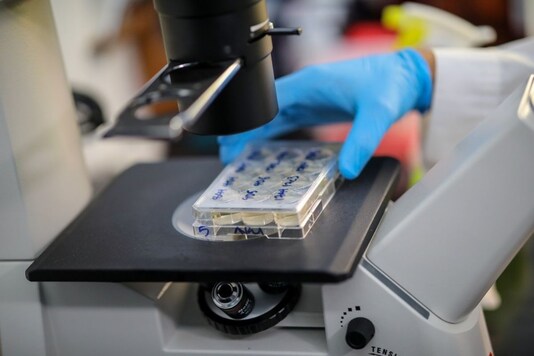
Scientists from Japan had a major breakthrough in the field of fluorescence microscopy recently when they developed a new technique to acquire fluorescence lifetime images. For the uninitiated fluorescence microscopy is a widely used technique in biochemistry and life sciences. It allows researchers to observe tiny cells and their surroundings.
The conventional techniques of fluorescence microscopy have a major limitation that its results are often difficult to quantitatively evaluate. However, the new technique developed by the scientists from the Institute of Post-LED Photonics, Tokushima University, leverages a phenomenon that is independent of experimental conditions. It helps quantify the fluorescent molecules.
According to the researchers, when a fluorescent material is subjected to radiation with a short burst of light, the fluorescence it produces does not immediately disappear but decays over a period of time, which varies from substance to substance.
The fluorescence decay is a fast process and is very difficult to capture with an ordinary camera. For this a single-point photo-detector is used that helps construct a complete 2-D image from each measured point. However, this method involves use of mechanical pieces which slows the imaging process.
In the new study, the researchers have developed a way to capture fluorescence lifetime images, without making use of the mechanical scanning.
“Our method can be interpreted as simultaneously mapping 44,400 light-based 'stopwatches' over a 2-D space to measure fluorescence lifetimes—all in a single shot and without scanning,” said Professor Takeshi Yasui of Tokushima University.
They use an optical frequency comb, which is a light signal made up of the sum of several discrete optical frequencies, as the excitation light for the sample. The term “comb” signifies how the signal looks when it is drawn against optical frequency. It looks like a dense cluster of equidistant spikes that resembles a hair comb.
The new microscopy method developed provides a high speed and spatial resolution, which will be very helpful in life sciences, especially where minute living cells are required to be observed.
Professor Yasui said that since the technique does not require scanning, a continuous measurement over the sample will be guaranteed in every shot. This approach will provide deeper insight into the numerous biological processes. It will also be useful in simultaneous imaging of multiple samples for testing antigens, something that is already in use to diagnose the COVID-19 virus.
The study highlights how optical frequency combs can be used in microscopy techniques which would help push the boundaries of innovation in the field of life sciences. It can facilitate the development of novel therapeutic techniques that can be employed for the treatment of intractable diseases. This in turn would help enhance the life expectancy of people.
