Mumbai: Keyhole surgey saves 87-year-old's life

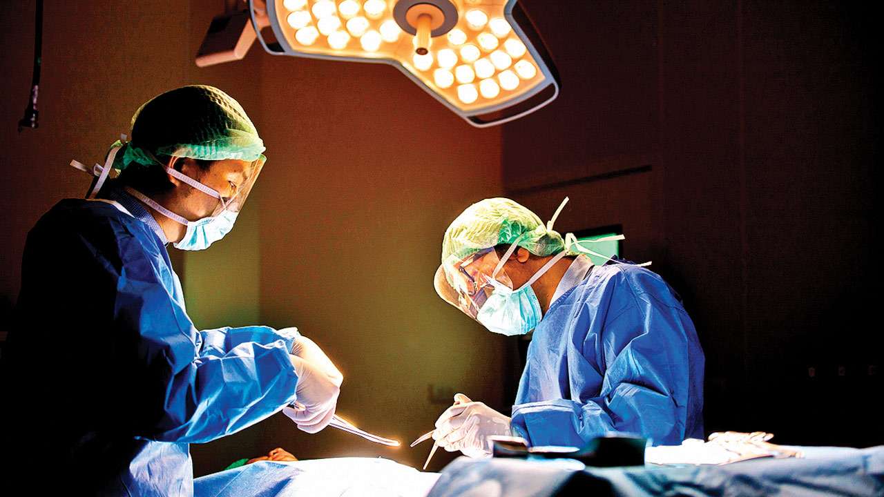
Surgery Picture for representation , Thinkstock
An 87-year-old man diagonised with thoracic and abdominal aortic aneurysm got a new lease of life after a team of four doctors at a city hospital used minimal invasive procedure to treat the octogenarian.
Hemraj Haria, a Wadala resident, had a mild abdominal pain when he came to the hospital where he was diagnosed with aortic aneurysm — ballooning or large dilation of both abdominal and thoracic regions of the aorta, which is the largest and main artery in the body. In case of a likely rupture, serious complications or instant death could have occurred.
"The aneurysm of the thoracic aorta was more than double the normal size," said Dr Bipeenchandra Bhamre, Cardiovascular surgeon, Sir HN Reliance Foundation Hospital, where Haria was admitted.
Given Haria's age, it was decided they'll treat the aneurysms with a minimally invasive procedure endovascular aortic stent grafting. In this technique, a graft is placed at the site of aneurysm by catheter technique avoiding the traditional open bypass surgery and the risks associated with it.
The doctors, however, faced a hurdle as this procedure requires usage of contrast agents to visualise the blood vessels. "During the investigations, we found that his kidney functions were slightly elevated. Using contrast agents could have further damaged the kidneys. Hence we opted for intra-vascular ultrasound imaging to help us visualise the vessels to properly position the graft. This reduced the need for contrast agents, helping reduce the risk of kidney damage," said Dr Rahul Sheth, who is an endovascular aortic specialist at the same hospital.
According to the hospital, this was the first time the procedure was employed in Western India, where the ultrasound imaging of the aorta was used to help accurately visualise the normal segments and place the device in the aorta to seal the aneurysm.
"He was admitted to the hospital on November 21 and was operated on the next day. While he was discharged yesterday (November 28), he needs to undergo another procedure for the abdominal aortic aneurysm after eight weeks time," said Mayur Haria, the patient's son.
UNIQUE PROCEDURE
This was the first time the procedure was employed in Western India where the ultrasound imaging of the aorta was used