


An Updated Review on: Liposomes as drug delivery system


 ABOUT AUHTORS
Devender Sharma*1, Aashiya Aara E. Ali 2, Leena R. Trivedi2
1Hi – Tech College of pharmacy, Chandrapur Maharashtra (India)
2Kamla Nehru College of pharmacy, Nagpur, Maharashtra (India)
*sdevender350@gmail.com
ABSTRACT
Liposomes are most placed acquiring in pharma industries and very useful in the various drug delivery system used to target the drug to particular tissue. Because of structural similarity between lipid bilayer (two layer) and cell membrane, liposome can easily penetrate effectively deliver drug to such that a free drug would not easily penetrate. Liposomes can be also encapsulate in both hydrophilic and hydrophobic materials, and are utilized as drug carriers in drug delivery. This technology is very useful for the treatment of certain diseases. Now a day’s most of the researcher attraction and interest will increase for that technology i.e. Liposomes. Main object of this review this technology i.e. Liposomes very useful in certain disease and easily prepare and also give various advantages other than. Liposomes are highly biocompatible, with applications ranging from delivering enzymes, antibacterial, antiviral drugs, antiparasite drugs, transdermal transporters, fungicides, diagnostic tools and adjuvant for vaccines. This paper mainly focus on exclusively scalable techniques and also focus on strength, respectively, limitations in respect to industrial applicability and regulatory requirements concerning liposomal drug formulations based on FDA and EMEA documents.
Reference Id: PHARMATUTOR-ART-2565
INTRODUCTION
Liposomes
Liposomes consist of vesicles composed of bilayers or multilayers that contain or have phospholipids and cholesterol surrounding an aqueous compartment. Drug is entrapped within the liposome and is released from the liposome for absorption at the intestinal membrane surface. This dosage form received considerable and this may well relate to their absorption enhancing ability, the feasibility of their use to promote drug absorption is uncertain drugs or chemical entities. Advances in combinatorial chemistry have led to the discovery of a wide number of new chemical entities (NCE) or drugs that have a potential therapeutic action on the biological systems. But most of the NCEs or drugs being discovered provide a challenge or produce most difficulties to the formulation scientist because of their physicochemical properties like poor solubility and permeability. Even though, above problems or difficulties could be addressed, but most of the molecules do not show or they fail their desired therapeutic action in vivo, which leads to lack of in vitro – in vivo correlation. A majority of anti-neoplastic agents, which are highly cytotoxicity to tumor cells in vitro, affect the normal cells also. This is due to their low therapeutic index (TI), i.e., the dose required to produce anti-tumor effect is toxic to normal cells. Such drugs have to be targeted to a specific site (diseased site) in order to reduce their toxic effects to normal tissues. Hence, an efficient drug delivery system is required to present the maximum fraction of administered dose at the target site or valuable for targeted sites. (Amidon et al., 1995)
Various carriers like nanoparticles, microparticles, polysaccharides, lectins and liposomes can be used to target the drug to a specific site. Liposomal drug delivery is gaining interest due to its contribution to varied areas like drug delivery, cosmetics, and structure of biological membrane. Liposomes very useful because act as a carrier for a variety of drugs, having a potential therapeutic action or other properties. Liposomes are colloidal carriers, having a size range of 0.01–5.0μm in diameter. Indeed these are bilayer vesicles that are formed when phospholipids are hydrated in excess of aqueous medium or aqueous solution. Liposomes have got a potential advantage of encapsulating hydrophilic as well as hydrophobic drugs and targeting them to the phospholipids are hydrated in excess of aqueous medium. Liposomes have got a potential advantage of encapsulating hydrophilic as well as hydrophobic drugs and targeting them to the required diseased site in the body. (Sunil et al., 2005)
Structure of liposome
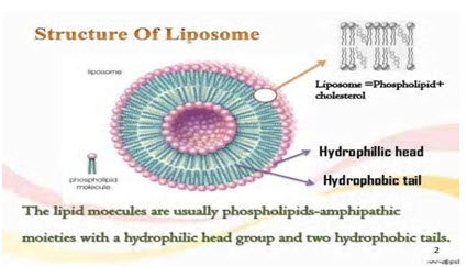
Table: Advantages and disadvantages of liposome (Sharma et al., 1997)
|
Advantages of liposome |
Disadvantages of liposome |
|---|---|
Stability increased if liposome prepared via encapsulation |
Short half-life
|
Liposomes increased efficacy and therapeutic index of drug (actinomycin-D) |
Low solubility |
Liposomes reduce the toxicity of the encapsulated agent (amphotericin B, Taxol) |
Leakage and fusion of encapsulated drug/molecules |
Liposomes help reduce the exposure of sensitive tissues to toxic drugs |
Production cost is high |
Site avoidance effect |
Fewer stables |
Liposomes are flexible, non-toxic, biocompatible, completely biodegradable, and non-immunogenic for systemic and non-systemic administrations |
Sometimes phospholipids undergoes oxidation and hydrolysis-like reaction |
Flexibility to couple with site-specific ligands to achieve active targeting |
|
Mechanism of liposome formation:
The basic or important part of liposome is formed by phospholipids, which are amphiphilic molecules (having a hydrophilic head and hydrophobic tail). The hydrophilic part is important it is mainly phosphoric acid bound to a water soluble molecule, whereas, the hydrophobic part consists of two fatty acid chains with 10-24 carbon atoms and 0-6 double bonds in each chain. When these phospholipids are disseminate or dispersed in aqueous medium, they form lamellar sheets by organizing in such a way that, the polar head group faces outwards to the aqueous region while the fatty acid groups face each other and finally form spherical/vesicle like structures called as liposomes. The polar portion remains or residue part in contact or touch with aqueous region along with shielding or keep safe of the non-polar part (which is oriented at an angle to the membrane surface). (Gregoriadis., 1979)
When phospholipids are hydrated in water, along with the input of energy like Sonication, shaking, heating, homogenization, etc. it is the hydrophilic/ hydrophobic interactions between lipid–lipid, lipid–water molecules that lead or show to the formation of bilayer vesicles in order to achieve or arrive at a thermodynamic equilibrium in the aqueous phase. (Gregoriadis., 1979) The main cause for bilayer formation includes:
1. The unfavorable interaction generated between hydrophilic and hydrophobic phase can be minimized or decreased by folding into closed concentric vesicles.
2. Large bilayer vesicle formation assists the reduction of large free energy difference present between the hydrophilic and hydrophobic environment.
3. Maximum stability to super molecular self assembled structure can be attained by forming into vesicles.
Classification of liposomes: (Gregoriadis., 1979)
Various classes of liposomes have been reported in various research and review paper. They are classified based on their size, number of bilayer, composition and method of preparation. Based on the size and number of bilayers, liposomes are classified as multilamellar vesicles (MLV), large unilamellar vesicles (LUV) and small unilamellar vesicles (SUV) as depicted in Fig. 2. Based on composition, they are classified as conventional liposomes (CL), pH-sensitive liposomes, cationic liposomes, long circulating liposomes (LCL) and immuno-liposomes. Based on the method of preparation, they are classified as reverse phase evaporation vesicles (REV), French press vesicles (FPV) and ether injection vesicles (EIV). In this context, the classification based on size and number of bilayer is discussed below. (Albertsson et al., 1985) (Donaruma et al., 1985)
Size range of liposome
|
Types of liposome |
Size range (nm) |
|
Small unilamellar vesicles |
20-40 |
|
Medium unilamellar vesicles |
40-80 |
|
Large unilamellar vesicles |
100-1000 |
Multilamellar vesicles (MLV)
MLV have a size greater than 0.1μm and consists of two or more bilayer. Their method of formulation is simple and very easy to carry which includes thin–film hydration method or hydration of lipids in excess of organic solvent. They are mechanically stable on long storage. Due to the large size, they are cleared early or rapidly by the reticulo-endithelial system (RES) cells and hence can be beneficial for various targeting the organs of RES. MLV have a moderate trapped volume, i.e., amount of aqueous volume to lipid ratio. The drug entrapment or corporate into the vesicles can be enhanced by slower rate of hydration and gentle mixing. Hydrating thin films of dry lipids can also easily enhance encapsulation efficiency. Subsequent lyophilization and rehydration after mixing with the aqueous phase (containing the drug) can yield MLV with good encapsulation efficiency i.e. 40%.
Large unilamellar vesicles (LUV) (Donaruma et al., 1985)
This class of liposomes particularly large unilamellar vesicles consists of a single bilayer and has a size greater than 0.1μm. They have higher encapsulation efficiency, since they can hold a large volume of solution in their cavity.
They have high trapped volume and can be useful for encapsulating hydrophilic drugs. Most useful advantage of LUV is that less amount of lipid is required for encapsulating large quantity of drug. Similar to MLV, they are rapidly cleared by RES cells, due to their larger size. LUV can be formulated by various methods like ether injection, detergent dialysis and reverse phase evaporation techniques. Apart from these methods, freeze thawing of liposomes, dehydration/ rehydration of SUV and slow swelling of lipids in non-electrolyte solution can a prepare LUV.
Small unilamellar vesicles (SUV) (Abra et al., 1981)
SUV are smaller in size (less than 0.1 μm) when compared to MLV and LUV, and have a single bilayer. They have a low entrapped aqueous volume to lipid ratio and characterized by having long circulation half life. SUV can be prepared by using solvent injection method (ethanol or ether injection methods) or alternatively by reducing the size of MLV or LUV using sonication or extrusion process under an inert atmosphere like nitrogen or Argon. The sonication can be performed using either a bath or probe type sonicator. SUV can also be achieved by passing MLV through a narrow orifice under high pressure. These SUV are susceptible to aggregation and fusion at lower or negligible/ no charge.
Methods of preparation: (Abra et al., 1981)
The conventional methods for preparing liposomes include solubilizing the lipids in organic solvent, drying down the lipids from organic solution, dispersion of lipids in aqueous media, purification of resultant liposomes and analysis of the final product. Of all the methods used for preparing liposomes, thin-film hydration method is the most simple and widely used one. MLV are produced by this method within a size range of 1–5μm. If the drug is hydrophilic it is included in the aqueous buffer and if the drug is hydrophobic, it can be included in the lipid film. But the drawback of this method is poor encapsulation efficiency (5-15% only) for hydrophobic drugs. By hydrating the lipids in presence of organic solvent, the encapsulation efficiency of the MLV can be increased. LUV can be prepared by solvent injection, detergent dialysis; calcium induced fusion and reverse phase evaporation techniques. SUV can be prepared by the extrusion or sonication of MLV or LUV. All these preparation methods involve the usage of organic solvents or detergents whose presence even in minute quantities can lead to toxicity. In order to avoid this, other methods like polyol dilution, bubble method and heating method have been developed without using any organic solvents or detergents. Detailed procedures for liposome preparation can be obtained from literature. (Tirrell et al., 1971) (Sahoo et al., 2003)
General methods of preparation (Gabizon et al., 1998)
All the methods of preparing the liposomes or formulating the liposomes involve four basic stages:
1. Drying down lipids from organic solvent.
2. Dispersing the lipid in aqueous media.
3. Purifying the resultant liposome.
4. Analyzing the final product.
Method of liposome preparation and drug loading (Abra et al., 1981)
The following methods are used for the preparation of liposome:
1. Passive loading techniques
2. Active loading technique.
Passive loading techniques include three different methods:
1. Mechanical dispersion method.
2. Solvent dispersion method.
3. Detergent removal method (removal of non encapsulated material)
Mechanical dispersion method (Tirrell et al., 1971)
The following are types of mechanical dispersion methods:
1.1. Sonication.
1.2. French pressure cell: extrusion.
1.3. Freeze-thawed liposomes.
1.4. Lipid film hydration by hand shaking, non-hand, shaking or freeze drying.
1.5. Micro-emulsification.
1.6. Membrane extrusion.
1.7. Dried reconstituted vesicles.
NOW YOU CAN ALSO PUBLISH YOUR ARTICLE ONLINE.
SUBMIT YOUR ARTICLE/PROJECT AT editor-in-chief@pharmatutor.org
Subscribe to Pharmatutor Alerts by Email
FIND OUT MORE ARTICLES AT OUR DATABASE
Recent Topics